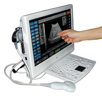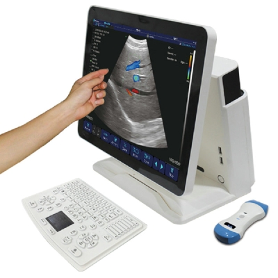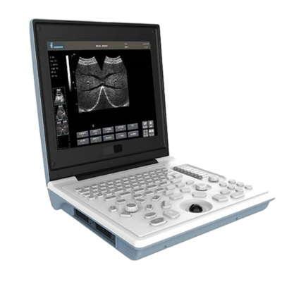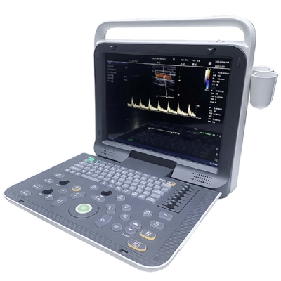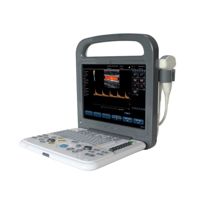
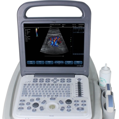
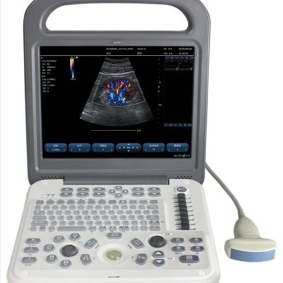
Specifications
| Technology platform | WINDOWS/PC platform |
| Probe interface | zero force metal body connector, effectively activated two mutual common interfaces |
| Frequency | 2.0-10MHz Variable frequency, frequency range 2.0-10MHz |
| Optional Probes | Convex, Linear, Transvaginal, Phased Array, Microconvex |
| Abdomen/Superficial/Cardiac/Transvaginal | 2.5-6.0MHz,5.0-10MHz,2.0-3.5MHz,5.0-9MHZ |
| B/D Dual-purpose | Linear: B/PW, Convex: B/PW, Sector-Scan: B/PW, Phased Array: B/PW |
| Imaging modes | B, 2B, 4B, M, Color, PDI, PW, CW, Dissection M, B+C+PW, B+PDI+PW |
| Imaging speed | Max 80 frames per second |
| Beam focus | 4 focal points |
| Real-time amplification | Unlimited |
| Freeze amplification | 3 times |
| Receiving mode | Parallel processing of multi-beam signals |
| Scanning Depth | ≥351mm |
| Playback | image playback for 48 seconds (Phased Array B mode) |
| False color | 7 kinds of color |
| Segment adjustment | 8 segments |
| Sector Scan Angle | 4 grades adjustable |
| 2D Modes(B) Phased Array | maximum: ≥6898 frames, Color,PDI maximum: ≥4050 frames |
| Spectral Doppler requirements | 1)Transmission Mode: pulse wave doppler PW, continuous wave doppler CW 2)PW testing range 0~7.5m/s 3)Doppler Frequency: PW frequency 4)Maximum measured velocity: positive or reverse flow velocity of 7.5m/s 5)Doppler Automatic envelope measurement and calculation 6)SV sampling width and location range : width 1-8mm 7)Display Control: reverse display (left/right;Up/down) 8)PW Real-time automatic measurement function 8)Scaleplate ≥16 grades; PRF 0.7kHz-9.3KHz adjustable 10)Playback: automatic playback of movies |
| System Imaging Function | 1)Color Doppler Enhancement Technology 2)Two-dimensional grayscale imaging 3)Power Doppler imaging 4)PHI pulse inverse phase tissue harmonic imaging + frequency composite technique 5)With the working mode of spatial composite imaging 6)Linear array probe independent deflection imaging technique 7)Linear trapezoidal spread imaging 8)B/Color/PW trisynchronous technology 9)Multibeam parallel processing 10)Speckle noise suppression technology 11)Convex expansion imaging 12)B-mode image enhancement technique 13)Logic view |
| Battery | Built-in large-capacity lithium battery, continuous working time ≥ 1.5 hours |
| General measurement | Distance, area, circumference, volume, angle, area ratio, distance ratio, angle, S/D velocity, time, heart rate, acceleration, etc |
| Input/output signal | Input: Mquipped with digital signal interface. Output: VGA, s-video,USB, audio interface, network interface. Connectivity: Medical digital imaging and communications DICOM3.0 interface components. Support network real-time transmission: can real-time transmission of user data to the server. Image management and recording device: 500G hard disk Ultrasonic image archiving and medical record management function: complete the storage management and playback storage of patient static image and dynamic image in the host computer |
| Color Doppler requirements | 1)The doppler gain is continuously adjustable 2)Color enhancement 3)B+COLOR display on both left and right sides of the same screen 4)Color mode baseline adjustment ±15 grades |